

Affecting up to 70–80% of women in their reproductive years, these common benign growths can cause heavy bleeding and pelvic pain, though are often asymptomatic and undiagnosed.
Understanding uterine fibroids
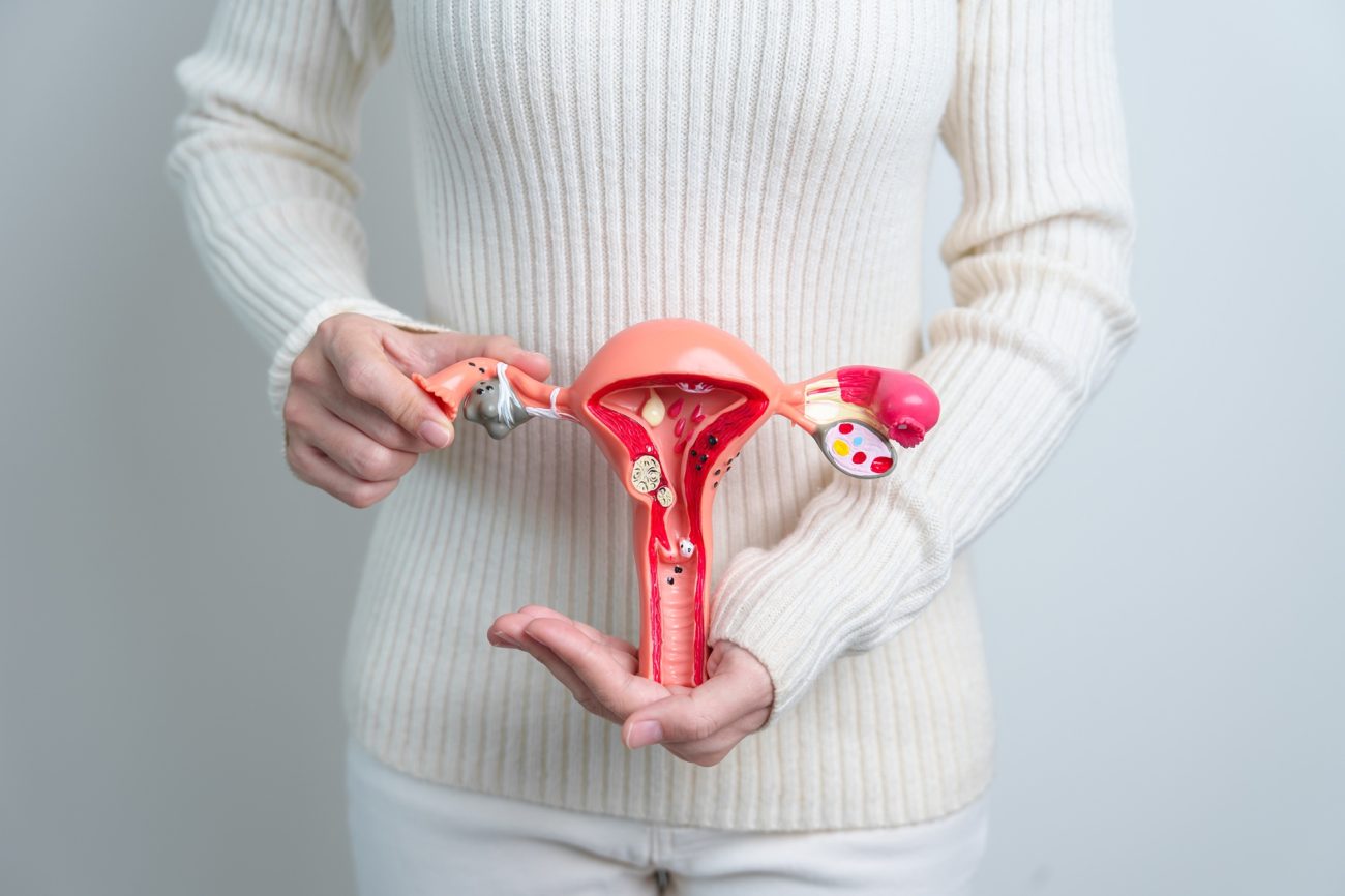
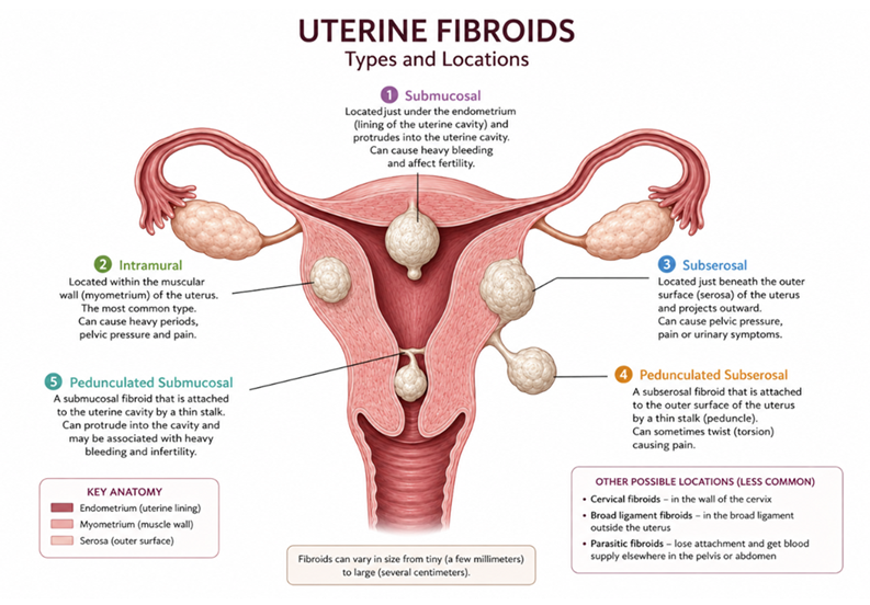
Uterine fibroids are a type of leiomyoma (benign tumours originating from smooth muscle cells) affecting up to 70–80% of women, most commonly during their reproductive years (25–45 years)(1,2). They can present anywhere in the uterine wall and are classified by location into submucosal, intramural and subserosal types (see Figure 1).
Size varies from a few millimetres to decimetres, historically measured in equivalent gestational uterine size (e.g., “12 weeks”) though volume or diameter is now the preferred measure (3).
The FIGO (International Federation of Gynecology and Obstetrics) classification further divides fibroid presentation into types 0–8 based on location (4). Diagnosis is generally made through abdominal palpation, ultrasound and MRI (magnetic resonance imaging) (2).
Around 30% of fibroids are symptomatic (2). Heavy menstrual bleeding is the most common presentation, and the resulting blood loss can cause large clots and iron-deficiency anaemia (3). Large fibroids may also press on surrounding tissue and organs, causing pain, dysfunction and a sensation of pelvic fullness or heaviness (1). Size and location both influence symptom severity, with large intramural fibroids in particular carrying a greater risk of fertility issues and miscarriage (2,4). Despite this symptom burden, fibroids remain predominantly benign, with only around a 0.2% risk of becoming malignant (cancerous) (3).
How do uterine fibroids work?
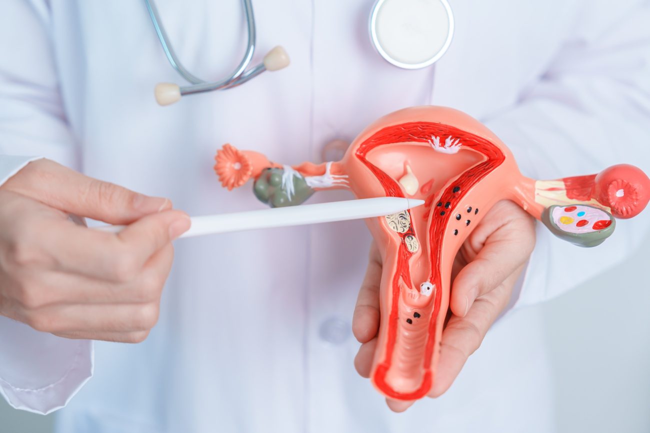
Uterine fibroids originate from a mutation in a single myometrial (uterine smooth muscle) stem cell. This is most commonly associated with genetic changes in the MED12 gene, present in around 70% of fibroids, among other implicated mutations (1,5,6). This neoplastic transformation proliferates, forming a benign tumour (1,6). The resulting fibroid is made up of these myometrial stem cells, differentiated fibroid smooth muscle cells, and tumour-associated fibroblasts, which produce the fibrous matrix that gives fibroids their name (1).
Fibroids are hormone-dependent, growing under the influence of oestrogen and progesterone and typically regressing postmenopausally (5). Oestrogen plays a priming role, upregulating oestrogen (ER) and progesterone receptors (PRs) on fibroid cells and increasing their sensitivity to both hormones (6,7). Progesterone is considered the primary driver of growth, acting through three mechanisms: suppressing apoptosis (programmed cell death), upregulating stem cell division, and increasing vascular endothelial growth factor (VEGF), promoting fibroid blood supply (7). This explains why fibroids can be problematic in pregnancy, tending to grow throughout gestation and raising the risk of miscarriage and complications (1,5).
Conventional management includes both medical and surgical options (8). Medical treatments include tranexamic acid, progestins, progesterone antagonists, GnRH agonists, and hormonal contraception; surgical interventions include myomectomy (fibroid removal), hysterectomy (removal of the uterus), and uterine artery embolisation (cutting the fibroid’s blood supply) (2,5). These interventions vary considerably in fertility preservation. Denosumab, a newer RANKL inhibitor, suppresses fibroid growth but is unsuitable in pregnancy due to effects on foetal bone development (9).
Understanding the root causes of uterine fibroids

While the disease mechanism is well understood, the epidemiology and associated risk factors for fibroids suggest the root causes are more complex. Overweight and obese women show an increased risk of developing fibroids (3). Other associated risk factors include race, nulliparity (having never carried a full-term pregnancy), vitamin D deficiency, exposure to endocrine-disrupting chemicals, hypertension (high blood pressure) and diabetes (5,8,10).
The obesity link, though not fully understood, is thought to involve increased conversion of androgens to oestrone (an oestrogen form) in adipose tissue, raising ER and PR expression and driving progression (11). Endocrine-disrupting chemicals (EDCs) are similarly thought to affect the hormone balance — mimicking endogenous hormones (made in the body), driving disease, and damaging DNA, causing mutations (10,12).
A 2003 US study found fibroids affect Black and Afro-Caribbean women disproportionately, with 80% prevalence and larger fibroid size at younger ages, compared to 70% prevalence in white Caucasian women (2,13). Notably, only 35% of the 1,364 women studied had a prior diagnosis, suggesting substantial historical underdiagnosis (2,13).
Vitamin D is a group of steroid compounds acting as hormones in the body, D3 (cholecalciferol) is synthesised in skin exposed to sunlight and obtained from dietary meat, fish and dairy (14). Vitamin D has shown anti-proliferative and pro-apoptotic effects, inducing cell differentiation and downregulating oestrogen and progesterone receptors across various diseases (14,15).
A 2013 study of 1,000 women found a significant correlation between vitamin D deficiency and fibroid presence, along with a reduction in fibroid mass following supplementation (15). However, a smaller 2020 study of 30 women found no significant change in fibroid size with supplementation (16). It has also been posited that the higher fibroid prevalence in Black women may relate to a greater sunlight requirement for adequate vitamin D synthesis in darker skin, alongside genetic factors (14,15,17,18).
The nulliparity association is notable, given that progesterone drives fibroid growth and levels are highest during pregnancy. However, postpartum uterine involution (shrinking) causes ischaemic breakdown of fibroid tissue — effectively a post-pregnancy “clean-up” that may offset fibroid growth during pregnancy (19).
Hypertension has also been identified as a key risk factor for fibroids. This is thought to relate to the shared smooth muscle origin of both tissues; fibroid cells express angiotensin II receptors, which mediate vasoconstriction in vascular smooth muscle via the renin-angiotensin-aldosterone system, potentially implicating this pathway in fibroid proliferation (20,21). Whether this reflects a causal pathway or two distinct co-existing smooth muscle diseases is unresolved. A large 2020 study (n=350,000) found a protective association between ACE (angiotensin-converting-enzyme) inhibitor use and fibroid incidence, supporting a shared pathway hypothesis (22).
Signs and symptoms
Common symptoms of uterine fibroids include:
- Heavy periods
- Long periods (<7 days)
- Pelvic pain
- Painful periods (dysmenorrhea)
- Lower back pain
- Urinary issues (frequency, urgency and bladder control)
- Constipation, bloating
- Painful bowel movements (dyschezia)
- Painful sex (dyspareunia)
- Difficulty getting pregnant
- Pregnancy loss or complications (1,2,8,23).
Less common symptoms:
- Iron-deficiency anaemia due to chronic blood loss
- Visible or palpable abdominal swelling
- Referred leg or hip pain due to nerve compression
- Acute pain from fibroid degeneration or torsion
- Venous thromboembolism
- Flank pain or kidney impairment due to ureteric compression (1,24,25,26,27,28)
When to seek urgent medical help
Emergency-level bleeding:
- Soaking through two maxi pads every hour for two or more consecutive hours
- Passing large clots repeatedly
- Bleeding accompanied by dizziness, fainting, rapid heartbeat, or shortness of breath — signs the body may be struggling to compensate for blood loss (23,24,26)
Sudden, severe pelvic pain:
Acute-onset pain, especially with fever, may indicate fibroid degeneration (the fibroid outgrowing its blood supply) or torsion of a pedunculated fibroid — both require urgent assessment (25,27).
Signs of venous thromboembolism:
Reported rarely, where large fibroids exert pressure on pelvic veins. Seek emergency care for:
- Unilateral leg swelling, pain, or warmth
- Sudden breathlessness, chest pain, or coughing up blood (28)
Any bleeding after menopause, regardless of volume, as other causes need to be ruled out (24,26).
Herbs for uterine fibroids
Anti-proliferative
- Green tea (Camellia sinensis) EGCG is the primary constituent. It is an antioxidant and has anti-angiogenic and anti-proliferative effects against fibroids (29,30).
- Turmeric (Curcuma longa) contains the constituent curcumin, shown to be anti-proliferative against multiple tumour cell lines, including fibroids (29,30).
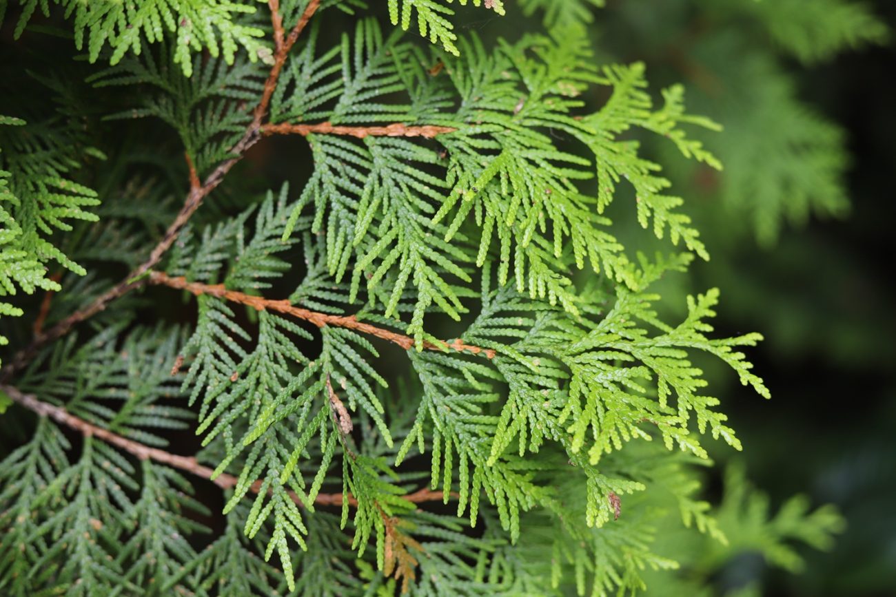
- Thuja (Thuja occidentalis) and greater celandine (Chelidonium majus) are anti-proliferative and used in the treatment of benign growths (31,32).
Excess oestrogen clearance
- Liver-supporting herbs like dandelion root (Taraxacum officinale), schisandra (Schisandra chinensis) and milk thistle (Silybum marianum) can improve excess hormone clearance (24,31,32).
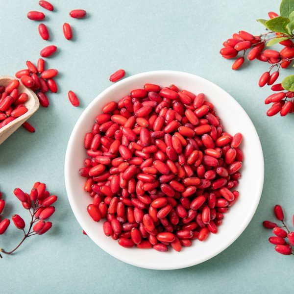
- Barberry (Berberis vulgaris) and greater celandine (Chelidonium majus) are cholagogues, stimulating bile production and the elimination of excess oestrogen in the gut (24,31).
Reducing menstrual bleeding
- Yarrow (Achillea millefolium) is effective in reducing both excess menstrual bleeding and dysmenorrhea (period pain) (31,33).
- Shepherd’s purse (Capsella bursa-pastoris) is used as a uterine anti-haemorrhagic (31,34).
- Lady’s mantle (Alchemilla vulgaris) is a uterine astringent and reduces blood loss (31,32).
- Agnus castus (Vitex agnus-castus) regulates the menstrual cycle, reducing prolactin levels and increasing dopamine in the pituitary (31,32). It is also used in the treatment of dysmenorrhea and excessive uterine bleeding (34).
Other support
- Nervines such as St John’s wort (Hypericum perforatum), lemon balm (Melissa officinalis) and motherwort (Leonurus cardiaca), and adaptogens like ashwagandha (Withania somnifera), can help reduce stress and improve hormone balance (32).
- Ginger (Zingiber officinale) reduces inflammation (32).
- Herbs like dang gui (Angelica sinensis), cinnamon (Cinnamomum cassia) and bai shao (Paeonia lactiflora) improve pelvic circulation and reduce “blood stasis” (24).
Holistic solutions
Diet
Vegetables and fibre
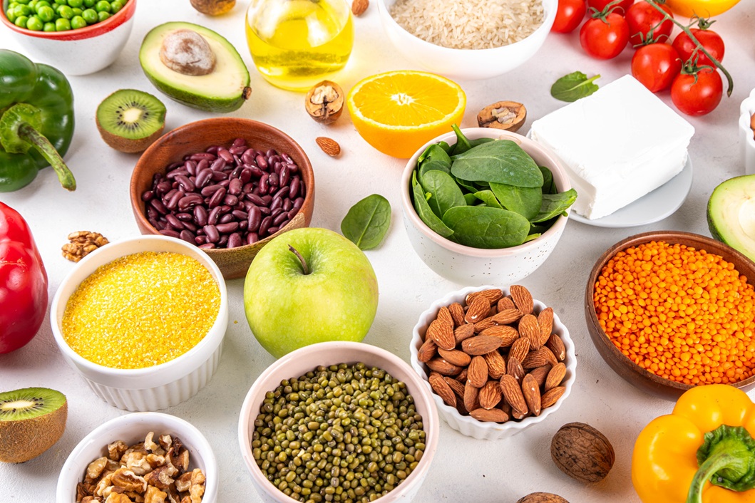
A diet rich in vegetables and fibre appears protective against uterine fibroids, by promoting favourable estrogen metabolism and reducing inflammation (30,35). Fibre aids in the excretion of excess estrogen from the body, lowering the risk of estrogen-driven fibroid growth (36). Cruciferous vegetables are particularly beneficial; they contain sulforaphane, a compound that supports healthy estrogen metabolism (37).
Omega-3
Higher dietary omega-3 polyunsaturated fatty acid intake has been associated with significantly reduced fibroid risk (30). Omega-3s are anti-inflammatory and key to cell plasma membrane integrity; they may also downregulate genes involved in fibroid cell function (29,30).
Vitamin D supplementation
Supplementation reduces fibroid volume and surgical intervention rates (30). Serum levels >30 ng/mL correlate with decreased fibroid incidence, while levels >20 ng/mL reduce fibroid growth (29). In a study, increased dairy intake correlated with decreased fibroid risk due to vitamin D content (38).
Limit or avoid
- Red meat: High red meat consumption is a potential risk factor for uterine fibroids, likely through its contribution to elevated circulating estrogen levels via saturated fat content (30). Women with or at risk of fibroids may benefit from limiting red meat intake as part of a dietary approach to manage this condition (29).
- High sugar diet: Research suggests high dietary glycemic load is associated with increased fibroid risk in younger women, potentially through elevated insulin and bioavailable estrogen, and increased inflammation (30,39).
Lifestyle factors
Endocrine-disrupting chemicals, particularly phthalates, bisphenol A, and diethylstilbestrol, affect pathways relevant to fibroid progression by mimicking the body’s hormones and causing epigenetic changes, driving mutation (12). Avoiding endocrine disruptors by limiting perfumes, plastic use and receipt handling, could help protect against developing fibroids (10,12).
Exercise has been linked to a reduced incidence of fibroid development, whilst a sedentary lifestyle correlates to a doubled risk (40,41).
References
- Bulun SE, Yin P, Wei JJ, et al. Uterine fibroids. Physiological reviews. 2025;105(4):10.1152/physrev.00010.2024. https://doi.org/10.1152/physrev.00010.2024
- Giuliani E, As‐Sanie S, Marsh EE. Epidemiology and management of uterine fibroids. International Journal of Gynecology & Obstetrics. 2020;149(1):3-9. https://doi.org/10.1002/ijgo.13102
- British Fibroid Trust. Fibroids: causes – types – symptoms. Www.britishfibroidtrust.org.uk. Published 2021. Accessed July 8, 2026. http://www.britishfibroidtrust.org.uk/Fib_info/wif.php
- Micić J, Macura M, Andjić M, et al. Currently available treatment modalities for uterine fibroids. Medicina. 2024;60(6):868-868. https://doi.org/10.3390/medicina60060868
- Saini K, Singh S, Garg N. Progesterone and estrogen signaling in uterine fibroids: A tale of two interconnected pathways. The Journal of Steroid Biochemistry and Molecular Biology. 2026;262:107036. https://doi.org/10.1016/j.jsbmb.2026.107036
- Stewart EA, Nowak RA. Uterine fibroids: hiding in plain sight. Physiology. 2022;37(1):16-27. https://doi.org/10.1152/physiol.00013.2021
- Ali M, Ciebiera M, Vafaei S, et al. Progesterone signaling and uterine fibroid pathogenesis; molecular mechanisms and potential therapeutics. Cells. 2023;12(8):1117-1117. https://doi.org/10.3390/cells12081117
- NHS. Overview – Fibroids. NHS. Published 2019. Accessed July 8, 2026. https://www.nhs.uk/conditions/fibroids/
- Ikhena DE, Liu S, Kujawa S, et al. RANKL/RANK pathway and its inhibitor RANK-Fc in uterine leiomyoma growth. The Journal of Clinical Endocrinology & Metabolism. 2018;103(5):1842-1849. https://doi.org/10.1210/jc.2017-01585
- Stewart E, Cookson C, Gandolfo R, Schulze-Rath R. Epidemiology of uterine fibroids: A systematic review. BJOG: An International Journal of Obstetrics & Gynaecology. 2017;124(10):1501-1512. https://doi.org/10.1111/1471-0528.14640
- Qin H, Lin Z, Vásquez E, Luan X, Guo F, Xu L. Association between obesity and the risk of uterine fibroids: A systematic review and meta-analysis. Journal of Epidemiology and Community Health. Published online October 16, 2020:jech-2019-213364. https://doi.org/10.1136/jech-2019-213364
- Bariani MV, Rangaswamy R, Siblini H, Yang Q, Al-Hendy A, Zota AR. The role of endocrine-disrupting chemicals in uterine fibroid pathogenesis. Current Opinion in Endocrinology, Diabetes & Obesity. 2020;27(6):380-387. https://doi.org/10.1097/med.0000000000000578
- Day Baird D, Dunson DB, Hill MC, Cousins D, Schectman JM. High cumulative incidence of uterine leiomyoma in black and white women: Ultrasound evidence. American Journal of Obstetrics and Gynecology. 2003;188(1):100-107. https://doi.org/10.1067/mob.2003.99
- Ciebiera M, Ali M, Prince L, et al. The evolving role of natural compounds in the medical treatment of uterine fibroids. Journal of Clinical Medicine. 2020;9(5). https://doi.org/10.3390/jcm9051479
- Baird DD, Hill MC, Schectman JM, Hollis BW. Vitamin D and the risk of uterine fibroids. Epidemiology. 2013;24(3):447-453. https://doi.org/10.1097/ede.0b013e31828acca0
- Arjeh S, Darsareh F, Asl ZA, Azizi Kutenaei M. Effect of oral consumption of vitamin D on uterine fibroids: A randomized clinical trial. Complementary Therapies in Clinical Practice. 2020;39:101159. https://doi.org/10.1016/j.ctcp.2020.101159
- Pavone D, Clemenza S, Sorbi F, Fambrini M, Petraglia F. Epidemiology and risk factors of uterine fibroids. Best Practice & Research Clinical Obstetrics & Gynaecology. 2018;46:3-11. https://doi.org/10.1016/j.bpobgyn.2017.09.004
- Williams J, Forlenza K, Swede H, Luciano D, Ulrich A. Vitamin D deficiency in black women with uterine fibroids. American Journal of Obstetrics and Gynecology. 2024;230(4):S1185-S1186. https://doi.org/10.1016/j.ajog.2024.02.072
- Laughlin SK, Hartmann KE, Baird DD. Postpartum factors and natural fibroid regression. American Journal of Obstetrics and Gynecology. 2011;204(6):496.e1-496.e6. https://doi.org/10.1016/j.ajog.2011.02.018
- Kirschen GW, AlAshqar A, Miyashita-Ishiwata M, Reschke L, El Sabeh M, Borahay MA. Vascular biology of uterine fibroids: Connecting fibroids and vascular disorders. Reproduction. 2021;162(2). https://doi.org/10.1530/rep-21-0087
- Stewart EA, Borah BJ. Uterine fibroids and hypertension: Steps toward understanding the link. The Journal of Clinical Endocrinology & Metabolism. 2020;106(2):e1039-e1041. Accessed July 12, 2026. https://academic.oup.com/jcem/article/106/2/e1039/5974965?login=false
- Fischer NM, Nieuwenhuis TO, Singh B, Yenokyan G, Segars JH. Angiotensin-converting enzyme inhibitors reduce uterine fibroid incidence in hypertensive women. The Journal of clinical endocrinology and metabolism. 2021;106(2):e650-e659. https://doi.org/10.1210/clinem/dgaa718
- Munro MG, Critchley HOD, Fraser IS. The two FIGO systems for normal and abnormal uterine bleeding symptoms and classification of causes of abnormal uterine bleeding in the reproductive years: 2018 Revisions. International Journal of Gynecology & Obstetrics. 2018;143(3):393-408. https://doi.org/10.1002/ijgo.12666
- Romm A. Botanical Medicine for Women’s Health. London: Elsevier Health Sciences; 2017.
- Charles K, Raoul K, Idrissa G, Diomande G, Serge B. Torsion of uterine fibroid: A rare cause of acute pelvic pain: About one case. Gynecology & Obstetrics Case report. 2017;03(03). https://doi.org/10.21767/2471-8165.1000056
- NICE. Recommendations | Heavy menstrual bleeding: Assessment and management | Guidance | NICE. Nice.org.uk. Published March 14, 2018. Accessed July 12, 2026. https://www.nice.org.uk/guidance/ng88/chapter/recommendations
- Samanta A, Bera R, Barua M, Debbarma D, Pande A. Acute uterine torsion masquerading as fibroid degeneration in a high-risk pregnancy: A case of diagnostic surprise. Cureus. Published online July 2, 2025. https://doi.org/10.7759/cureus.87156
- Sparić R, Stojković M, Šarac M, et al. Venous thromboembolism associated with uterine fibroids: A review of reported cases. Journal of Clinical Medicine. 2026;15(2):444. https://doi.org/10.3390/jcm15020444
- Krzyżanowski J, Paszkowski T, Woźniak S. The Role of nutrition in pathogenesis of uterine fibroids. Nutrients. 2023;15(23):4984-4984. https://doi.org/10.3390/nu15234984
- Martire FG, Costantini E, Ianes I, et al. Nutrition and uterine fibroids: Clinical impact and emerging therapeutic perspectives. Journal of Clinical Medicine. 2025;14(20):7140. https://doi.org/10.3390/jcm14207140
- Brice-Ytsma H, McDermott A. Herbal Medicine in Treating Gynaecological Conditions. Aeon Books; 2020.
- Reilly M. Herbal Medicine and Reproductive Health. Aeon Books; 2021.
- Moradi M, Niazi A, Abbasi K, Mollazadeh S, Feizabadi M. The impact of achillea millefolium on primary dysmenorrhea and menstrual bleeding: A systematic review. Journal of Midwifery and Reproductive Health. 2024;12(2). https://doi.org/10.22038/JMRH.2023.68062.1991
- Bone K, Mills S. Principles and Practice of Phytotherapy: Modern Herbal Medicine. 2nd ed. Edinburgh: Churchill Livingstone, Elsevier; 2013.
- Islam MS, Segars JH, Castellucci M, Ciarmela P. Dietary phytochemicals for possible preventive and therapeutic option of uterine fibroids: Signaling pathways as target. Pharmacological Reports. 2017;69(1):57-70. https://doi.org/10.1016/j.pharep.2016.10.013
- Szydłowska I, Nawrocka-Rutkowska J, Brodowska A, Marciniak A, Starczewski A, Szczuko M. Dietary natural compounds and vitamins as potential cofactors in uterine fibroids growth and development. Nutrients. 2022;14(4):734. https://doi.org/10.3390/nu14040734
- Islam MS, Cayton Vaught KC, Brennan JT, Segars JH. Sulforaphane, an organic isothiocyanate found in cruciferous vegetables, regulates expression of genes involved in hippo signaling and fibrotic phenotype in uterine fibroid cells. Fertility and Sterility. 2021;116(3):e318. https://doi.org/10.1016/j.fertnstert.2021.07.856
- Wise LA, Radin RG, Palmer JR, Kumanyika SK, Rosenberg L. A prospective study of dairy intake and risk of uterine leiomyomata. American Journal of Epidemiology. 2009;171(2):221-232. https://doi.org/10.1093/aje/kwp355
- Radin RG, Palmer JR, Rosenberg L, Kumanyika SK, Wise LA. Dietary glycemic index and load in relation to risk of uterine leiomyomata in the Black Women’s Health Study. The American Journal of Clinical Nutrition. 2010;91(5):1281-1288. https://doi.org/10.3945/ajcn.2009.28698
- Baird DD, Dunson DB, Hill MC, Cousins D, Schectman JM. Association of physical activity with development of uterine leiomyoma. American Journal of Epidemiology. 2007;165(2):157-163. https://doi.org/10.1093/aje/kwj363
- British Medical Journal. 6+ Hours/day of sedentary leisure time linked to doubling in fibroids risk – BMJ Group. BMJ Group – Helping doctors make better decisions. Published November 29, 2023. Accessed July 14, 2026. https://bmjgroup.com/6-hours-day-of-sedentary-leisure-time-linked-to-doubling-in-fibroids-risk/