







Genetic, immune and microbiome mechanisms driving endometriosis are informing new diagnostics, conventional research and herbal approaches to inflammation and hormonal balance.
An endometriosis recap
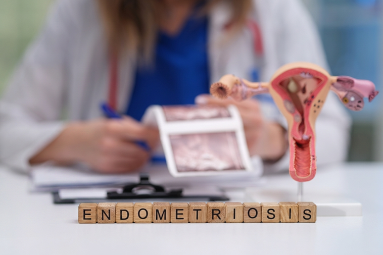
Endometriosis is a chronic inflammatory condition in which tissue similar to the lining of the uterus (the endometrium) forms lesions outside the usual place. Most settle within the pelvis, but they have been documented as far afield as the chest and, rarely, the brain (1,2,3). Characteristic symptoms include dysmenorrhoea (painful periods), chronic pelvic pain, dyschezia (painful bowel movements), dysuria (painful urination), dyspareunia (painful sex), fatigue and infertility (1,2). See our article on endometriosis for a full summary.
Despite affecting roughly 10% of reproductive-aged women, endometriosis has long been poorly understood. Often framed as a disease of retrograde menstruation (backflow of menstrual blood into the pelvis) compounded by oestrogen excess, its underlying mechanisms are now becoming clearer (3). Newer research reframes it as a systemic inflammatory condition marked by significant immune dysfunction, and is beginning to establish the microbiome’s role in lesion development and oestrogen dominance (4,5).
Diagnosis takes an average of seven to nine years and was historically possible only via laparoscopy (keyhole surgery) (1,6). Since 2022, first-line investigation has been ultrasound and MRI, though imaging cannot rule out mild disease, and no reliable diagnostic blood test yet exists (1,6).
As of 2026, this may be changing: two new non-invasive tests have provisionally joined NICE guidance for a three-year evidence-gathering period (7). Endotest is a microRNA saliva test, whilst Endosure uses electrical mapping of the digestive tract to detect signalling changes caused by endometriosis; together they could significantly cut diagnosis times, costs and the need for invasive surgery (7,8,9). Endosure also generates scores that may help monitor disease severity and treatment response, though this is not yet confirmed (9).
Conventional treatment remains non-curative: NSAIDs, progestins and other hormonal medications manage symptoms but inhibit pregnancy (1). Surgical removal of lesions and adhesions (internal scarring) — considered the gold standard — carries a recurrence risk and can itself cause further scarring and reduced fertility (6).
For a disease associated with an estimated 40% of female infertility, 60% of chronic pelvic pain, burdened by long diagnostic delays and non-curative treatment options, what new insights are emerging into how endometriosis works — and what role might herbal medicine play in supporting those affected (6)?
Genetic links
Endometriosis runs in families; close relatives carry a roughly 6–15% risk of disease, yet no single gene explains it (10,11). Genome-wide association studies (GWAS), which scan the whole genome for variants linked to a condition, have identified around 80 associated regions, none appearing consistently across cases (12).
Many of these variants are shared with other pain-related and inflammatory conditions; coeliac disease; for instance, occurs around three times more often alongside endometriosis (12,13). The genes implicated govern hormone balance, immune function, inflammation, immune evasion and tissue invasion — the same mechanisms research finds integral to how the disease develops (12).
Immune cell dysfunction

Retrograde menstruation occurs in around 90% of women, yet only about 10% develop endometriosis. This points to a disease origin in a failure of immune-system clearance (3). Across both, its innate (immediate) and adaptive (learned) arms, immune function is not only impaired, but reprogrammed to support ectopic lesions, reflecting similarities to the way tumours evade immune attack (3,14,15).
Macrophages are immune cells responsible for the removal and digestion of debris, pathogens and faulty cells. In early disease, macrophages display a pro-inflammatory (M1) form, releasing chemical messengers that drive inflammation (3,14,16). Increased red blood cells in the pelvis during menstruation overload their capacity for phagocytosis (engulfing), inhibiting their ability to clear unwanted cells (15). As disease progresses, they shift to an anti-inflammatory M2 type, which secretes different types of chemical messengers (IL-10, TGF-β, and VEGF), driving scarring and angiogenesis (new blood vessel formation) (3,14,15).
Neutrophils are cytotoxic (cell-killing) cells that help clear endometrial tissue during menstruation and trap microbes by casting web-like neutrophil extracellular traps (NETs) (16). In endometriosis, NETs are overproduced, hinting at infective involvement (14,16). A landmark 2026 study went further, identifying a neutrophil subset in patients’ pelvic fluid, primed to release NETs that physically capture endometrial cells and drive new lesion growth (17).
The trigger traced back to gut bacteria (particularly Pseudomonas) leaking across a compromised intestinal barrier into the pelvis, where their endotoxin sets off the NETs; degrading the NETs or blocking their formation suppressed lesions in mice (17). Neutrophils also help release the egg at ovulation, and failure of this (luteinised-unruptured follicle syndrome) is five times more common in endometriosis (13,18).
Natural killer (NK) cells, which are responsible for the removal of faulty or infected cells, become less cytotoxic, letting lesions persist (3,16). Mast cells cluster around lesions and release nerve and blood-vessel growth factors (NGF and VEGF), contributing to both pain and lesion growth (14).
The adaptive immune system also exhibits dysfunction. Locally, T-cells shift towards an anti-inflammatory, immunosuppressive profile that suppresses lesion clearance (Th2/Tregs); whilst systemically, they tip towards inflammation linked to failed embryo implantation (Th1), which is why immune-modulating treatments aimed at Th1/Th2 balance are being explored for endometriosis patients experiencing infertility (13,16). Finally, B-cells, which produce antibodies, are over-stimulated similarly to in autoimmune disease, generating autoantibodies against the body’s own tissue (19). Whilst endometriosis is not classified as autoimmune, this may help explain why it so often travels alongside autoimmune conditions (16,19).
Oestrogen dominance, the estrobolome and the microbiome

In endometriosis, lesions generate their own oestrogen through raised activity of aromatase (the enzyme that converts androgens into oestrogen), whilst becoming resistant to opposition by progesterone (3). This creates a local oestrogen dominance that feeds the immune dysfunction already described, promoting the M2 and Th2 shifts and mast-cell activation (15). The inflammatory pathways this generates (NF-κB and COX-2/PGE2) drive aromatase in turn, causing a self-reinforcing loop (3,15).
The microbiome has also been implicated in endometriosis. A community of gut bacteria known as the estrobolome produces β-glucuronidase, an enzyme that reactivates oestrogens the liver had tagged for excretion — returning them to circulation and raising systemic oestrogen levels (20,21,22). When the gut and reproductive-tract populations fall out of balance, protective Lactobacillus declines as unfavourable E. coli and Streptococcus rise, feeding this cycle of oestrogen recirculation, inflammation and immune evasion (22,23).
This dysbiosis connects back to the NETs described earlier. A healthy intestinal wall keeps bacteria contained; dysbiosis inflames and loosens those junctions (“leaky gut”), letting bacterial toxins (LPS) and even whole bacteria escape into the circulation and pelvis, driving systemic inflammation (17,21,23). LPS acts through a specific immune receptor, TLR4, switching on the same inflammatory NF-κB signalling, so the same gut permeability that triggers NETs also feeds the aromatase loop (20,22,23). Endometriosis patients show raised intestinal permeability and circulating LPS, with higher levels correlating to more severe disease; together with the Pseudomonas-linked NETs above, this builds a compelling case that dysbiosis fuels endometriosis (17,22,24,25).
There are still uncertainties in the microbiome link. A 2023 mouse study found Fusobacterium in the uterus could drive endometrial cells to become migratory and lesion-promoting, while antibiotics shrank lesions (4); yet the largest human study to date found no clear microbiome difference between affected and unaffected women (26). Gut and vaginal dysbiosis remain incompletely understood, but point to practical approaches, from restoring the gut barrier to modulating the microbiome (24).
Holistic approaches to endometriosis

Microbiome
The microbiome comes in here too: Other commensal (beneficial) gut bacteria ferment fibre into short-chain fatty acids (SCFAs) such as butyrate, which calm inflammatory NF-κB signalling and have been shown to suppress endometriotic cell survival (21,27). SCFAs can heal gut permeability, reducing local gut inflammation and the release of endotoxins into systemic circulation (21,24,27). This offers a mechanistic rationale for how a high-fibre diet could support endometriosis (21,24).
Targeting the microbiome directly is a significant new avenue. Two small trials found Lactobacillus probiotics reduced period pain versus placebo, though interventions ran only eight weeks, and in one, pain returned after the probiotic stopped, suggesting commensal levels receded (28,29,30). The first double-blind RCT designed specifically to test whether a probiotic can favourably modulate the estrobolome in endometriosis is now underway, and should offer firmer evidence (31). Given probiotics carry no established risk, they may be worth trying in the meantime (29,30).
Diet
Recent systematic reviews find the Mediterranean diet, including vegetables, fruit, legumes, oily fish and olive oil, strongly associated with reduced endometriosis symptoms, owing to the anti-inflammatory, antioxidant-rich polyphenol profile of these foods (32,33,34). Omega-3 fatty acids shift prostaglandin production away from the inflammatory loop that feeds oestrogen dominance (32,33,34,35), while dietary fibre feeds the bacteria that make the protective SCFAs covered earlier (21,32,33). Conversely, red meat and trans fats supply arachidonic acid, which may raise inflammation, and are associated with increased risk (32,33,34).
Sulforaphane, from cruciferous vegetables, exhibits antioxidant and anti-inflammatory effects, improving oestrogen detoxification in the liver and countering the NF-κB signalling seen in endometriosis (36,37). Human endometriosis-specific data is lacking, but the mechanistic case for including cabbage and broccoli is strong, with no associated risk (36,37).
Gluten-free and low-FODMAP diets showed variable results and poor adherence to strict elimination, so are best considered on an individual basis (32,33,34,35).
Other interventions
Unsurprisingly, regular physical activity has been linked to improved quality of life and reduced associated symptoms in a meta-analysis, exhibiting a lowering effect on systemic inflammation and circulating oestrogen levels (34,38). Additionally, exercise positively correlates to increased bone density, in loss due to GnRH agonist treatment (38,39).
Reducing exposure to endocrine-disrupting chemicals (EDCs) such as BPA and phthalates lowers intake of exogenous (from outside the body) oestrogens, which is supported by documented links between EDC exposure and endometriosis (40,41). Some research suggests these may be a significant contributing factor to disease onset (35).
New directions in conventional endometriosis treatment

Novel research looking at alternative treatments for endometriosis is focusing on targeting inflammation and immune evasion; some preliminary research is exploring immunotherapies targeting B and T-cell dysfunction (19,35). Other research is looking at prescribing off-label medications such as angiotensin-receptor blockers and certain biologics, aimed at inhibiting VEGF-mediated blood supply (35,42,43).
Many compounds under research derive from medicinal plants: silymarin from milk thistle (Silybum marianum) decreases inflammatory cytokines and reduces endometriotic ovarian cysts (35,44), while curcumin from turmeric (Curcuma longa) inhibits the inflammatory NF-κB pathway and is an antioxidant, though an RCT found no significant effect on pain or quality of life — possibly due to poor gut absorption (35,45,46).
Resveratrol (found in berries and nuts) is a potent antioxidant shown to inhibit NF-κB signalling and new blood vessel growth to lesions, though its bioavailability is poor (35,47,48). One in vitro endometriosis study found polydatin — a resveratrol glycoside from Japanese knotweed (Reynoutria japonica) — has higher bioavailability and converts readily to resveratrol, though this has not been tested in patients (48).
Herbal medicine for endometriosis
Herbal treatment for endometriosis currently targets inflammation, pain and cramping, and hormonal regulation (49,50,51,52,53). Common anti-inflammatory herbs used are turmeric, liquorice (Glycyrrhiza glabra) and ginger (Zingiber officinale), whilst cramp bark (Viburnum opulus) and pasqueflower (Pulsatilla vulgaris) are commonly cited for reducing pain and uterine spasm (49,50,51,52,53). Hormonal regulation is supported by enhancing phase II liver pathways to tag oestrogen for excretion with herbs like milk thistle and schisandra (Schisandra chinensis) (50).
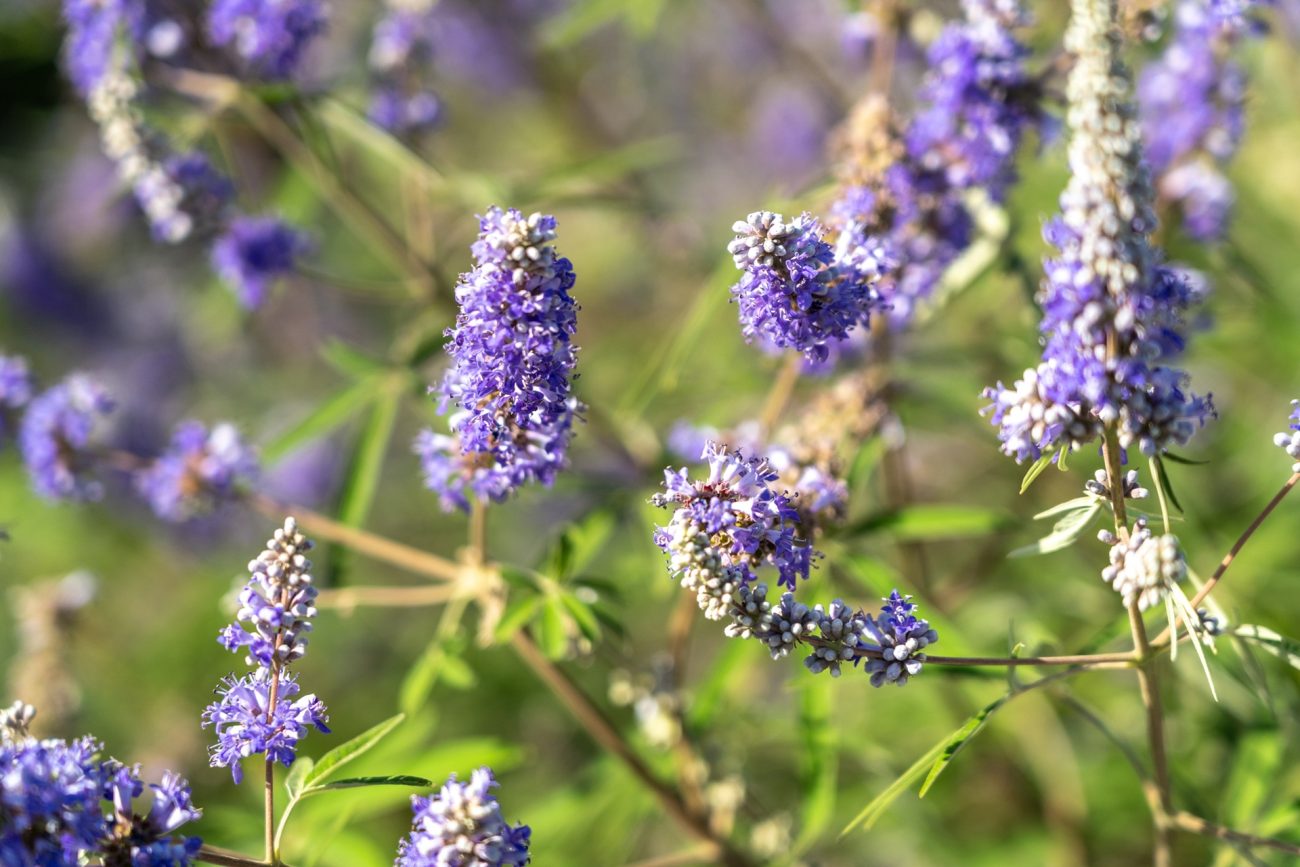
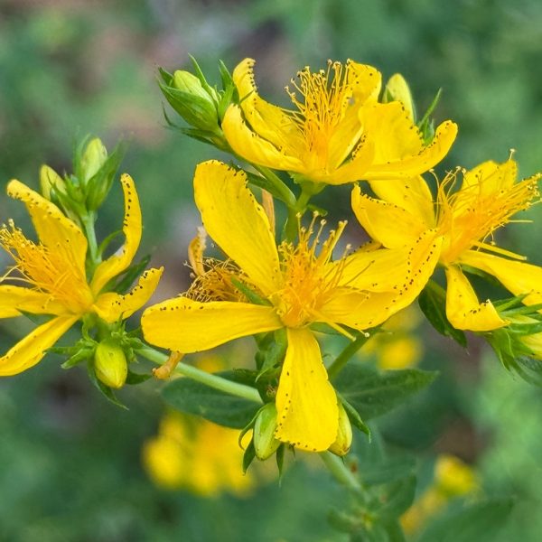
Vitex (Vitex agnus-castus) is regularly recommended for hormone regulation in endometriosis (49,50,51,52,53) and has evidence for period pain, though no endometriosis-specific studies exist (54,55). Its primary action is in the pituitary: diterpenes bind dopamine D2 receptors to inhibit prolactin, relieving its suppression of the corpus luteum and so raising post-ovulatory progesterone — an ill-fitting mechanism to the oestrogen dominance and progesterone resistance of endometriosis (56).
Some vitex constituents also bind oestrogen receptors, potentially counterproductive in an oestrogen-excess condition (55,57). More promisingly, in vitro research suggests β-sitosterol and casticin have aromatase-inhibitory activity, better aligned with the aromatase upregulation seen in endometriosis (55,58). On balance, vitex warrants case-by-case mechanistic consideration rather than routine use.
Given that gut inflammation and intestinal permeability contribute to endometriosis symptoms and progression, mucilage-containing herbs like plantain (Plantago major) and marshmallow root (Althaea officinalis), which can be fermented into SCFAs in the gut, may offer benefit (21,27,50).
The immune-dysfunction framing also makes a case for immune-modulating herbs: polysaccharides in medicinal mushrooms such as reishi (Ganoderma lucidum) and roots like echinacea (Echinacea angustifolia) can help rebalance the Th2/Th1 imbalance described earlier (16,59,60).
EGCG from green tea (Camellia sinensis) has been shown to reduce endometriosis lesions and inhibit VEGF in animal trials, whilst also exhibiting potent antioxidant capacity, reducing inflammation (44).
Endometriosis, with its complex mechanisms and treatment avenues, is a fast-moving field. Research into immune dysfunction, the microbiome and oestrogen metabolism is beginning to demystify the disease, though deeper understanding and better treatments are still needed. These mechanistic insights may point to new avenues for support — conventionally and in herbal medicine.
AI statement
AI was employed in the initial literature search stage as well as to order the bibliography. All work is the product of the author.
References
- European Society of Human Reproduction and Embryology. Endometriosis guideline. ESHRE website. Published 2022. Accessed July 27, 2026. https://www.eshre.eu/Guideline/Endometriosis
- National Institute for Health and Care Excellence. Endometriosis: diagnosis and management. NICE guideline NG73. Published 2017. Accessed July 30, 2026. https://www.nice.org.uk/guidance/ng73
- Shifon S, Tyrinova T, Veretelnikova T, Pasman N, Chernykh E. Endometriosis as an immune-mediated disease: pathogenetic mechanisms and therapeutic strategies. Front Immunol. 2025;16. https://doi.org/10.3389/fimmu.2025.1727183
- Muraoka A, Suzuki M, Hamaguchi T, et al. Fusobacterium infection facilitates the development of endometriosis through the phenotypic transition of endometrial fibroblasts. Sci Transl Med. 2023;15(700). https://doi.org/10.1126/scitranslmed.add1531
- Li W, Feng H, Ye Q. Factors contributing to the delayed diagnosis of endometriosis—a systematic review and meta-analysis. Front Med. 2025;12. https://doi.org/10.3389/fmed.2025.1576490
- Sardo A, Becker CM, Renner SP, et al. Management of women with endometriosis in the 21st century. Curr Opin Obstet Gynecol. 2025;37(3):149-157. https://doi.org/10.1097/gco.0000000000001027
- National Institute for Health and Care Excellence. New technologies could help cut years-long wait for endometriosis diagnosis, says NICE. NICE website. Published July 7, 2026. Accessed July 24, 2026. https://www.nice.org.uk/news/articles/new-technologies-for-endometriosis-diagnosis-in-primary-care
- Ziwig. Discover Ziwig Endotest. Ziwig website. Published 2016. Accessed July 27, 2026. https://ziwig.com/en/discover-ziwig-endotest/
- EndoSure. EndoSure homepage. EndoSure website. Published 2022. Accessed July 24, 2026. https://www.endosure.com/
- Guo P, Gan J, Xu L, Li W. Causal links and mediating effects of lipid metabolism, immune cells, and inflammatory proteins in endometriosis: evidence from Mendelian randomization. Medicine (Baltimore). 2025;104(28):e43163. https://doi.org/10.1097/md.0000000000043163
- Koninckx PR, Ussia A, Adamyan L, Wattiez A, Gomel V, Martin DC. Pathogenesis of endometriosis: the genetic/epigenetic theory. Fertil Steril. 2019;111(2):327-340. https://doi.org/10.1016/j.fertnstert.2018.10.013
- Koller D, He J, Løkhammer S, et al. Multi-ancestry genome-wide association and integrated multi-omics analyses of endometriosis and its clinical manifestations. Nat Genet. 2026;58(5):1-11. https://doi.org/10.1038/s41588-026-02582-2
- Maksym RB, Hoffmann-Młodzianowska M, Skibińska M, Rabijewski M, Mackiewicz A, Kieda C. Immunology and immunotherapy of endometriosis. J Clin Med. 2021;10(24):5879. https://doi.org/10.3390/jcm10245879
- Ahmed RS, Sherif M, Alghamdi MA, et al. Exploring the immune system’s role in endometriosis: insights into pathogenesis, pain, and treatment. Cureus. 2025;17(7):e87091. https://doi.org/10.7759/cureus.87091
- Zakiyah M, Asmarinah A. Mechanism of immune system dysfunction, apoptosis and oxidative stress on endometriosis. J Biomed Transl Res. 2023;9(2):88-95. https://doi.org/10.14710/jbtr.v9i2.16885
- Abramiuk M, Grywalska E, Małkowska P, Sierawska O, Hrynkiewicz R, Niedźwiedzka-Rystwej P. The role of the immune system in the development of endometriosis. Cells. 2022;11(13):2028. https://doi.org/10.3390/cells11132028
- Wu X, Wu M, Li H, et al. Intraperitoneal translocation of gut microbiota induces NETosis and promotes endometriosis. Gut. 2026;75(7):1110-1122. https://doi.org/10.1136/gutjnl-2025-336185
- Makinoda S, Hirosaki N, Waseda T, et al. Granulocyte colony-stimulating factor (G-CSF) in the mechanism of human ovulation and its clinical usefulness. Curr Med Chem. 2008;15(6):604-613. https://doi.org/10.2174/092986708783769740
- Mohseni M, Saravi M, Oskouie I, Zolbin M. The immunology of endometriosis and the therapeutic potential of bispecific antibodies: a hypothesis. Avicenna J Med Biotechnol. 2025. https://doi.org/10.18502/ajs.v7i4.17468
- Ervin SM, Li H, Lim L, et al. Gut microbial β-glucuronidases reactivate estrogens as components of the estrobolome. J Biol Chem. 2019;294(49):18586-18599. https://doi.org/10.1074/jbc.RA119.010950
- Liang L, Min L, Liu J, Liu Y, Cheng W. Gut microbiota dysbiosis in endometriosis: mechanistic insights and gut microbiota-targeted therapeutic strategies. Front Microbiol. 2026;17. https://doi.org/10.3389/fmicb.2026.1776574
- Li Z, Yin Z, Chen W, Wang Z. Impact of gut and reproductive tract microbiota on estrogen metabolism in endometriosis. Am J Reprod Immunol. 2025;93(6). https://doi.org/10.1111/aji.70109
- Khan KN, Fujishita A, Hiraki K, et al. Bacterial contamination hypothesis: a new concept in endometriosis. Reprod Med Biol. 2018;17(2):125-133. https://doi.org/10.1002/rmb2.12083
- Viganó D, Zara F, Pinto S, et al. How is small bowel permeability in endometriosis patients? A case control pilot study. Gynecol Endocrinol. 2020;36(11):1010-1014. https://doi.org/10.1080/09513590.2020.1766440
- Xholli A, Cremonini F, Perugi I, Londero AP, Cagnacci A. Gut microbiota and endometriosis: exploring the relationship and therapeutic implications. Pharmaceuticals (Basel). 2023;16(12):1696. https://doi.org/10.3390/ph16121696
- Pérez-Prieto I, Vargas E, Salas-Espejo E, et al. Gut microbiome in endometriosis: a cohort study on 1000 individuals. BMC Med. 2024;22(1). https://doi.org/10.1186/s12916-024-03503-y
- Kim N, Yang C. Butyrate as a potential modulator in gynecological disease progression. Nutrients. 2024;16(23):4196. https://doi.org/10.3390/nu16234196
- Khodaverdi S, Mohammadbeigi R, Khaledi M, et al. Beneficial effects of oral Lactobacillus on pain severity in women suffering from endometriosis: a pilot placebo-controlled randomized clinical trial. Int J Fertil Steril. 2019;13(3):178-183. https://doi.org/10.22074/ijfs.2019.5584
- Itoh H, Uchida M, Sashihara T, et al. Lactobacillus gasseri OLL2809 is effective especially on the menstrual pain and dysmenorrhea in endometriosis patients: randomized, double-blind, placebo-controlled study. Cytotechnology. 2011;63(2):153-161. https://doi.org/10.1007/s10616-010-9326-5
- Hearn-Yeates F, Horne AW, O’Mahony SM, Saunders PTK. The impact of the microbiota-gut-brain axis on endometriosis-associated symptoms: mechanisms and opportunities for personalised management strategies. Reprod Fertil. 2024;5(2). https://doi.org/10.1530/raf-23-0085
- Kralj S, Zeman K, Mikuš M, et al. The role of probiotics in the treatment of endometriosis (ProMetrioS): a randomised double-blinded placebo-controlled cross-over trial. Trials. 2026;27(1). https://doi.org/10.1186/s13063-025-09405-5
- Nirgianakis K, Egger K, Kalaitzopoulos DR, Lanz S, Bally L, Mueller MD. Effectiveness of dietary interventions in the treatment of endometriosis: a systematic review. Reprod Sci. 2021;29(1). https://doi.org/10.1007/s43032-020-00418-w
- Neri L, Quintiero F, Fiorini S, et al. Diet and endometriosis: an umbrella review. Foods. 2025;14(12):2087. https://doi.org/10.3390/foods14122087
- Boroncsok D, Filó A, Török M, Vágó H, Ács N, Sobel G. The role of lifestyle and diet in the treatment of endometriosis: a review. Nutrients. 2026;18(1):142. https://doi.org/10.3390/nu18010142
- Ramos-Nino ME. Non-hormonal strategies in endometriosis: targets with future clinical potential. J Clin Med. 2025;14(14):5091. https://doi.org/10.3390/jcm14145091
- Fahey JW, Raphaely M. The impact of sulforaphane on sex-specific conditions and hormone balance: a comprehensive review. Appl Sci. 2025;15(2):522. https://doi.org/10.3390/app15020522
- Zhou A, Hong Y, Lv Y. Sulforaphane attenuates endometriosis in rat models through inhibiting PI3K/Akt signalling pathway. Dose Response. 2019;17(2):1559325819855538. https://doi.org/10.1177/1559325819855538
- Xie M, Qing X, Huang H, et al. The effectiveness and safety of physical activity and exercise on women with endometriosis: a systematic review and meta-analysis. PLoS One. 2025;20(2):e0317820. https://doi.org/10.1371/journal.pone.0317820
- Bergström I, Freyschuss B, Jacobsson H, Landgren BM. The effect of physical training on bone mineral density in women with endometriosis treated with GnRH analogs: a pilot study. Acta Obstet Gynecol Scand. 2005;84(4):380-383. https://doi.org/10.1111/j.0001-6349.2005.00558.x
- Ribeiro B, Mariana M, Lorigo M, Oliani D, Ramalhinho AC, Cairrão E. Association between the exposure to phthalates and the risk of endometriosis: an updated review. Biomedicines. 2024;12(8):1932. https://doi.org/10.3390/biomedicines12081932
- Wieczorek K, Szczęsna D, Jurewicz J. Environmental exposure to non-persistent endocrine disrupting chemicals and endometriosis: a systematic review. Int J Environ Res Public Health. 2022;19(9):5608. https://doi.org/10.3390/ijerph19095608
- Rocha A, Reis FM, Taylor RN. Angiogenesis and endometriosis. Obstet Gynecol Int. 2013;2013:1-8. https://doi.org/10.1155/2013/859619
- Moazen S, Arjmand MH. The role of local angiotensin II/angiotensin type 1 receptor in endometriosis: a potential target for new treatment approaches. Curr Mol Pharmacol. 2024;17. https://doi.org/10.2174/0118761429315431240712100124
- Mirzaei N, Jahanian Sadatmahalleh S, Rouholamin S, Nasiri M. A randomised trial assessing the efficacy of silymarin on endometrioma-related manifestations. Sci Rep. 2022;12(1). https://doi.org/10.1038/s41598-022-22073-8
- Vallée A, Lecarpentier Y. Curcumin and endometriosis. Int J Mol Sci. 2020;21(7). https://doi.org/10.3390/ijms21072440
- Gudarzi R, Shabani F, Charandabi S, Naghshineh E, Shaseb E, Mirghafourvand M. Effect of curcumin on painful symptoms of endometriosis: a triple-blind randomized controlled trial. Phytother Res. 2023. https://doi.org/10.1002/ptr.8030
- Jiang T, Chen Y, Gu X, et al. Review of the potential therapeutic effects and molecular mechanisms of resveratrol on endometriosis. Int J Womens Health. 2023;15:741-763. https://doi.org/10.2147/ijwh.s404660
- Gołąbek-Grenda A, Juzwa W, Kaczmarek M, Olejnik A. Resveratrol and its natural analogs mitigate immune dysregulation and oxidative imbalance in the endometriosis niche simulated in a co-culture system of endometriotic cells and macrophages. Nutrients. 2024;16(20):3483. https://doi.org/10.3390/nu16203483
- Romm A. Botanical Medicine for Women’s Health. Elsevier Health Sciences; 2018.
- Bone K, Mills S. Principles and Practice of Phytotherapy. Elsevier Health Sciences; 2013.
- Reilly M. Herbal Medicine and Reproductive Health. Aeon Books; 2021.
- Trickey R. Women, Hormones and the Menstrual Cycle. Allen & Unwin; 2011.
- Brice-Ytsma H, Chidley N. Herbal Medicine in Treating Gynaecological Conditions, Volume 2. Aeon Books; 2024.
- Höller M, Steindl H, Abramov-Sommariva D, et al. Use of Vitex agnus-castus in patients with menstrual cycle disorders: a single-center retrospective longitudinal cohort study. Arch Gynecol Obstet. 2024. https://doi.org/10.1007/s00404-023-07363-4
- Sirotkin AV. Effects, mechanisms of action and application of Vitex agnus-castus for improvement of health and female reproduction. Phytother Res. 2025;39(3):1484-1493. https://doi.org/10.1002/ptr.8438
- Puglia L, Lowry JV, Tamagno G. Vitex agnus-castus effects on hyperprolactinaemia. Front Endocrinol. 2023;14. https://doi.org/10.3389/fendo.2023.1269781
- European Medicines Agency. Assessment report on Vitex agnus-castus L., fructus. EMA website. Published 2018. Accessed July 29, 2026. https://www.ema.europa.eu/en/documents/herbal-report/final-assessment-report-vitex-agnus-castus-l-fructus-revision-1_en.pdf
- Dawood HM, Shawky E, Hammoda HM, Metwally AM, Ibrahim RS. Chemical constituents from Artemisia annua and Vitex agnus-castus as new aromatase inhibitors: in-vitro and in-silico studies. J Mex Chem Soc. 2020;64(4). https://doi.org/10.29356/jmcs.v64i4.1236
- Powell M. Medicinal Mushrooms: A Clinical Guide. Mycology Press; 2015.
- Ganora L. Herbal Constituents: Foundations of Phytochemistry. Herbalchem Press; 2021.